Development of an automated diagnostic system for malaria and urogenital schistosomiasis using artificial intelligence tools and a low-cost universal robotic microscope
Jun 04, 2025
Carles Rubio Maturana defended on June 3, his doctoral thesis on the diagnosis of malaria and urogenital schistosomiasis using artificial intelligence tools and a low-cost robotic microscope in the Doctoral Program in Microbiology of the Autonomous University of Barcelona under the direction of doctors Joan Joseph Munné and Elisa Sayrol Clols. The thesis is part of a project funded by the World Health Organization and directed by professor Daniel Lopez Codina from the Department of Physics at the UPC.
Malaria is one of the most prevalent infectious diseases in sub-Saharan Africa, with 263 million cases reported worldwide in 2023 according to the World Health Organization (WHO). Schistosomiasis is classified by the WHO as a neglected tropical disease, with more than 253 million people at risk of infection. Microscopy remains the gold standard technique for the diagnosis of both diseases. However, it is a professional-dependent method with a high level of involvement in the daily routine of laboratories. As an alternative, new diagnostic techniques based on image analysis with Artificial Intelligence (AI) tools are being developed.
Artificial Intelligence is one of the most disruptive emerging technologies of the current century, which has boosted and improved traditional methods of image analysis. Deep learning and the use of convolutional neural networks (CNN) for object detection in images and videos could be a suitable alternative to conventional microscopy diagnosis.
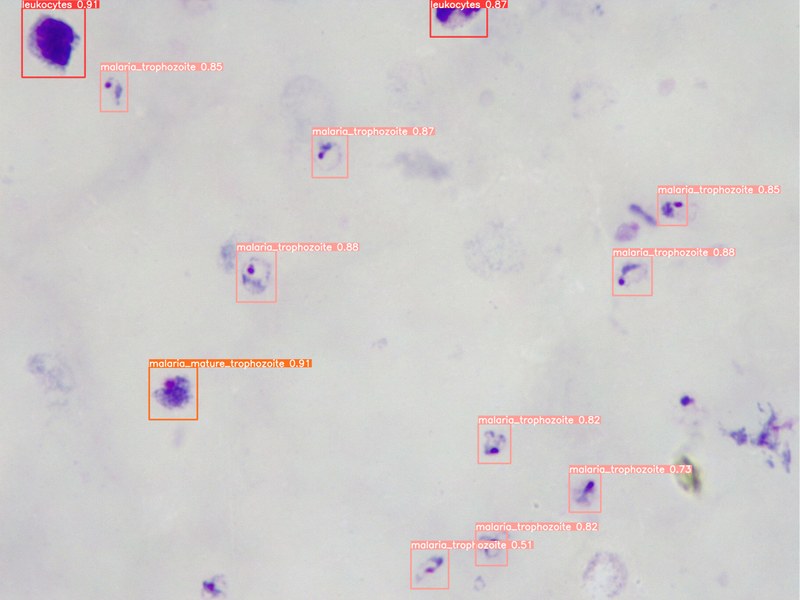
The diagnostic algorithms were integrated into a smartphone application and laboratory software designed for its handling on a computer. The system is capable of performing a fully automated diagnosis using: image autofocus and movements in the X-Y axes of the robotic microscope, CNN models trained for digital image analysis and the smartphone. The new prototype is capable of determining whether a Giemsa-stained thick drop sample is positive/negative for Plasmodium infection, and its parasitemia levels; detecting the presence of S. haematobium eggs and erythrocytes/leukocytes in images of urinary sediment. The results obtained in the validation at the Drassanes Vall d’Hebron Center demonstrated a sensitivity of 81.25% and a specificity of 92.11% for the diagnosis of malaria. On the other hand, a proof of concept has been carried out at the Nossa Senhora da Paz Hospital (Cubal, Angola) for the diagnosis of malaria, demonstrating satisfactory results for the implementation of the system in laboratories with few resources.

Share: